
📖 Step 2 — Learning Material
🔹 1️⃣ Introduction
The small intestine is the major site for digestion and absorption of nutrients.
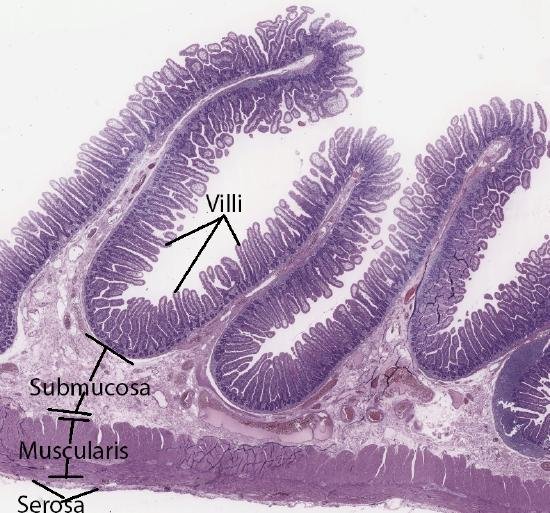
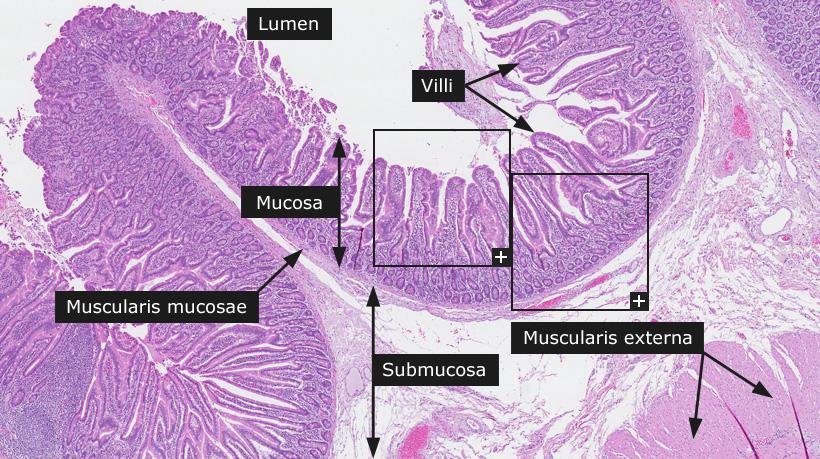
Its wall is specially adapted to increase surface area through plicae circulares, villi, and microvilli.
Histologically, the jejunum and ileum show important differences that help in identification under microscope.
The jejunum is mainly specialized for absorption, while the ileum also has an important immune role.
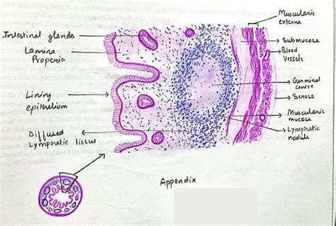
The appendix is a narrow lymphoid-rich part of the large intestine attached to the cecum.
Its histology is important because it explains the tendency of the appendix to inflammation and obstruction.
Understanding these microscopic features helps students connect structure with function and clinical disease.
🔹 2️⃣ Foundation Concepts
Key Definitions
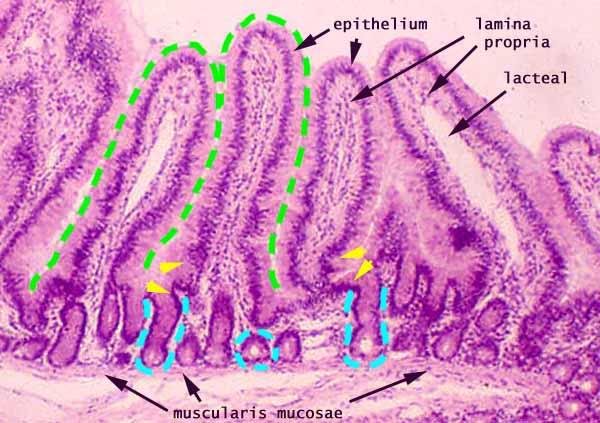
- Mucosa: Innermost layer of intestine containing epithelium, lamina propria, and muscularis mucosae.
- Submucosa: Connective tissue layer containing blood vessels, lymphatics, and nerve plexus.
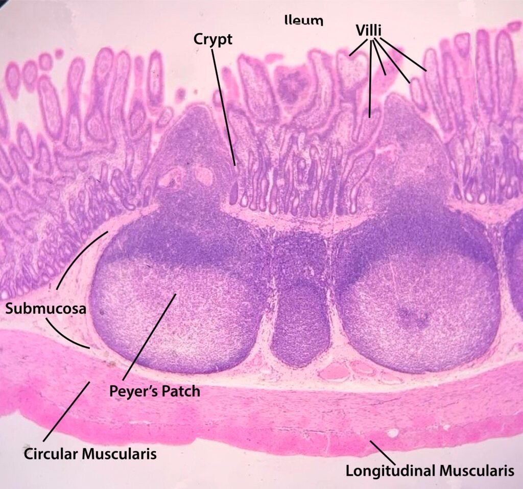
- Muscularis externa: Smooth muscle layer responsible for intestinal movement.
- Serosa: Outer covering formed by visceral peritoneum.
- Plicae circulares: Circular folds of mucosa and submucosa.
- Villi: Finger-like mucosal projections.
- Microvilli: Tiny projections on absorptive epithelial cells forming brush border.
- Crypts of Lieberkühn: Tubular intestinal glands opening between villi.
- Peyer’s patches: Aggregated lymphoid nodules mainly present in ileum.
- Appendix: Blind-ended lymphoid-rich tube attached to cecum.
🔹 3️⃣ Core Learning — Curriculum Coverage
1: Histology of Jejunum
🧠 CORE
- Jejunum is the middle part of the small intestine.
- It is mainly specialized for absorption of nutrients.
- It has tall, closely packed villi.
- Plicae circulares are large and prominent.
- Goblet cells are present but fewer than in ileum.
- Crypts of Lieberkühn are present between villi.
- Peyer’s patches are usually absent or poorly developed.
- Submucosa does not contain Brunner’s glands.
- Main recognition feature: tall villi + prominent plicae circulares.
🔬 CONCEPT EXPLAINED
The jejunum has a mucosa lined by simple columnar epithelium.
The epithelial cells are mainly absorptive enterocytes with microvilli forming a brush border.
The villi are long and finger-like. These increase the surface area available for absorption.
The plicae circulares are large folds containing mucosa and submucosa. They slow intestinal contents and allow more contact time for absorption.
Each villus contains lamina propria with blood capillaries and a central lacteal.
Capillaries absorb amino acids and sugars, while lacteals absorb fats.
Structure → Function:
Tall villi + prominent plicae circulares → increased surface area → efficient nutrient absorption.
⚠️ IF DAMAGED
Damage to jejunal mucosa causes loss of villi.
Cause → Effect:
- Villous atrophy → reduced surface area
- Reduced absorption → diarrhea and malnutrition
- Brush border damage → impaired digestion of nutrients
- Severe mucosal injury → weight loss and nutrient deficiency
2: Histology of Ileum
🧠 CORE
- Ileum is the distal part of the small intestine.
- It has shorter villi than jejunum.
- Plicae circulares are smaller and less prominent.
- Goblet cells are more numerous.
- Peyer’s patches are prominent in lamina propria and submucosa.
- Crypts of Lieberkühn are present.
- Ileum absorbs bile salts and vitamin B12.
- Main recognition feature: Peyer’s patches + shorter villi.
- It has both absorptive and immune functions.
🔬 CONCEPT EXPLAINED
The ileum is lined by simple columnar epithelium with absorptive cells and goblet cells.
Compared with jejunum, the villi are shorter and fewer.
The most important microscopic feature of ileum is the presence of Peyer’s patches.
These are large collections of lymphoid tissue present mainly on the antimesenteric border.
Goblet cells increase toward the distal intestine. They secrete mucus, which lubricates intestinal contents and protects the mucosa.
Structure → Function:
Peyer’s patches → immune surveillance → protection against intestinal pathogens.
⚠️ IF DAMAGED
Damage to ileal mucosa affects absorption and immunity.
Cause → Effect:
- Ileal disease → reduced bile salt absorption
- Bile salt loss → fat malabsorption
- Terminal ileal damage → vitamin B12 deficiency
- Peyer’s patch involvement → impaired intestinal immune defense
3: Plicae Circulares
🧠 CORE
- Plicae circulares are permanent circular folds.
- They are formed by mucosa and submucosa.
- They are most prominent in jejunum.
- They increase absorptive surface area.
- They slow the movement of chyme.
- They are absent in the appendix.
- They are different from villi because villi involve only mucosa.
- Main function: increase contact between chyme and mucosa.
🔬 CONCEPT EXPLAINED
Plicae circulares are large folds projecting into the intestinal lumen.
Because they include both mucosa and submucosa, they are larger and more permanent than villi.
They create a spiral movement of chyme. This allows nutrients to remain in contact with the absorptive surface for longer time.
They are highly developed in jejunum because jejunum is the major absorptive region.
Structure → Function:
Large circular folds → slow chyme movement → more contact time → better absorption.
⚠️ IF DAMAGED
Flattening or loss of folds reduces intestinal absorptive efficiency.
Cause → Effect:
- Loss of plicae → reduced surface area
- Rapid passage of chyme → poor absorption
- Malabsorption → diarrhea, weakness, weight loss
4: Crypts of Lieberkühn
🧠 CORE
- Crypts of Lieberkühn are tubular intestinal glands.
- They open between villi.
- They extend down into lamina propria.
- They contain stem cells for epithelial renewal.
- They contain goblet cells.
- They may contain Paneth cells.
- They help in secretion and epithelial replacement.
- They maintain the intestinal lining.
🔬 CONCEPT EXPLAINED
Crypts of Lieberkühn are simple tubular glands located between villi.
They are important because the intestinal epithelium is constantly exposed to enzymes, acid, bile, bacteria, and mechanical stress.
Stem cells in the crypts replace damaged epithelial cells.
Goblet cells secrete mucus.
Paneth cells help protect against bacteria.
Structure → Function:
Crypts with stem cells → epithelial renewal → healthy absorptive surface.
⚠️ IF DAMAGED
Damage to crypts prevents epithelial repair.
Cause → Effect:
- Stem cell injury → failure of mucosal renewal
- Mucosal breakdown → ulceration
- Loss of secretion → poor protection
- Increased infection and inflammation
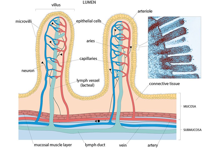
5: Villi
🧠 CORE
- Villi are finger-like projections of mucosa.
- They are lined by simple columnar epithelium.
- They contain lamina propria.
- Each villus contains blood capillaries.
- Each villus contains a central lacteal.
- Villi increase surface area.
- Jejunum has tall villi.
- Ileum has shorter villi.
- Villi are absent in appendix.
🔬 CONCEPT EXPLAINED
Villi project into the lumen and increase the absorptive surface.
Their epithelium contains absorptive enterocytes and goblet cells.
Inside each villus, capillaries absorb glucose and amino acids.
The central lacteal absorbs lipids in the form of chylomicrons.
Structure → Function:
Villus core with vessels + absorptive epithelium → rapid nutrient uptake.
⚠️ IF DAMAGED
Villous damage leads to malabsorption.
Cause → Effect:
- Villi become flattened → reduced surface area
- Nutrient absorption decreases → diarrhea
- Fat absorption decreases → steatorrhea
- Severe damage → weight loss and anemia
6: Microvilli
🧠 CORE
- Microvilli are tiny projections on enterocytes.
- They form the brush border.
- They greatly increase surface area.
- They contain digestive enzymes.
- They help in final digestion and absorption.
- They are not visible clearly by light microscope individually.
- Brush border may be seen as a fuzzy apical border.
- Main function: terminal digestion and absorption.
🔬 CONCEPT EXPLAINED
Microvilli are microscopic projections from the apical surface of absorptive cells.
Together they form the brush border.
The brush border contains enzymes that complete digestion of carbohydrates and proteins.
It also contains transport proteins that move nutrients into epithelial cells.
Structure → Function:
Microvilli + enzymes + transporters → final digestion → nutrient absorption.
⚠️ IF DAMAGED
Microvilli damage reduces brush border function.
Cause → Effect:
- Loss of brush border enzymes → incomplete digestion
- Reduced transporter function → poor absorption
- Osmotic load in lumen → diarrhea
7: Histology of Appendix
🧠 CORE
- Appendix is attached to cecum.
- It resembles large intestine but has abundant lymphoid tissue.
- It has mucosa lined by simple columnar epithelium.
- Villi are absent.
- Crypts are present but often shorter and fewer.
- Goblet cells are present.
- Lymphoid follicles are abundant in mucosa and submucosa.
- Muscularis externa is present.
- Main recognition feature: no villi + abundant lymphoid follicles.
🔬 CONCEPT EXPLAINED
The appendix has a narrow lumen and a wall rich in lymphoid tissue.
Unlike small intestine, it does not have villi because absorption is not its main function.
Its mucosa contains crypts of Lieberkühn and goblet cells.
The lymphoid follicles are very prominent and may surround much of the lumen.
The appendix plays a minor immune role, especially earlier in life.
Structure → Function:
Abundant lymphoid tissue → immune surveillance near cecum.
⚠️ IF DAMAGED
The narrow lumen of appendix can become obstructed.
Cause → Effect:
- Lymphoid swelling or fecalith → luminal obstruction
- Obstruction → bacterial overgrowth
- Increased pressure → inflammation
- Result → acute appendicitis
8: Peyer’s Patches
🧠 CORE
- Peyer’s patches are aggregated lymphoid nodules.
- They are mainly found in ileum.
- They are present in lamina propria and submucosa.
- They form part of gut-associated lymphoid tissue.
- They help monitor intestinal antigens.
- They protect against pathogens.
- They may interrupt villi in the involved region.
- Main recognition feature of ileum.
🔬 CONCEPT EXPLAINED
Peyer’s patches are collections of lymphocytes arranged as lymphoid nodules.
They are especially important in the ileum because the distal small intestine contains many microorganisms and antigens.
They sample antigens from the intestinal lumen and help initiate immune responses.
This protects the intestinal wall from infection.
Structure → Function:
Lymphoid nodules near mucosa → antigen detection → immune protection.
⚠️ IF DAMAGED
If Peyer’s patches are damaged, mucosal immune defense decreases.
Cause → Effect:
- Reduced lymphoid defense → increased infection risk
- Inflammation of lymphoid tissue → swelling
- Swelling near appendix/ileocecal region → possible luminal narrowing
9: Surface Area Specializations
🧠 CORE
The small intestine increases surface area at three levels:
- Plicae circulares
- Villi
- Microvilli
These specializations allow maximum absorption.
- Plicae circulares are gross mucosal-submucosal folds.
- Villi are microscopic mucosal projections.
- Microvilli are cell surface projections.
- Together they greatly increase absorptive capacity.
- They are most developed where absorption is highest.
🔬 CONCEPT EXPLAINED
The small intestine must absorb large amounts of nutrients in a limited length.
To solve this, its wall uses multiple levels of folding.
Plicae circulares increase the internal surface.
Villi add finger-like projections.
Microvilli form a brush border on enterocytes.
Structure → Function:
More folding → more surface area → more enzymes and transporters → better absorption.
⚠️ IF DAMAGED
Loss of surface area specializations causes malabsorption.
Cause → Effect:
- Villous flattening → decreased absorption
- Microvilli damage → reduced brush border enzymes
- Poor nutrient uptake → diarrhea and nutritional deficiency
⚙️ 4️⃣ Functional Flow
| Structure | Function | Outcome |
|---|---|---|
| Plicae circulares | Increase surface area and slow chyme | Better absorption |
| Villi | Provide absorptive projections | Rapid nutrient uptake |
| Microvilli | Brush border digestion and absorption | Final digestion completed |
| Crypts of Lieberkühn | Secretion and epithelial renewal | Maintains mucosal lining |
| Jejunum | Major absorption site | Nutrient absorption |
| Ileum | Absorption + immune defense | B12/bile salt absorption and protection |
| Peyer’s patches | Immune surveillance | Defense against pathogens |
| Appendix lymphoid tissue | Local immune role | Immune monitoring near cecum |
🩺 5️⃣ Clinical Correlation
1. Villous Atrophy
- Loss or flattening of villi reduces absorptive surface area.
- Causes malabsorption, diarrhea, weight loss, and nutrient deficiency.
- Important histological concept in intestinal disease.
2. Ileal Disease
- Terminal ileum absorbs vitamin B12 and bile salts.
- Damage may cause B12 deficiency and fat malabsorption.
- Students should remember ileum is both absorptive and immune.
3. Acute Appendicitis
- Appendix has a narrow lumen and abundant lymphoid tissue.
- Lymphoid swelling may obstruct the lumen.
- Obstruction leads to bacterial overgrowth and inflammation.
- Histology explains why appendix is prone to inflammation.
4. Peyer’s Patch Enlargement
- Peyer’s patches may enlarge during immune response.
- This can contribute to local swelling.
- Important recognition feature of ileum in histology slides.
📌 6️⃣ Summary Points
- Jejunum has tall villi and prominent plicae circulares.
- Ileum has shorter villi and prominent Peyer’s patches.
- Plicae circulares contain mucosa and submucosa.
- Villi contain lamina propria, capillaries, and central lacteal.
- Microvilli form the brush border of enterocytes.
- Crypts of Lieberkühn are intestinal glands between villi.
- Crypts contain stem cells for epithelial renewal.
- Peyer’s patches are aggregated lymphoid nodules of ileum.
- Appendix has no villi but has abundant lymphoid follicles.
- Main function of jejunum is absorption.
- Main special feature of ileum is immune surveillance.
- Appendicitis commonly results from obstruction of the narrow lumen.